Bilateral Pleural Effusion Ddx : Ppt Approach To Pleural Effusion Powerpoint Presentation Free Download Id 5879920 : Pleural effusion (transudate or exudate) is an accumulation of fluid in the chest or on the lung.
byAdmin•
0
Bilateral Pleural Effusion Ddx : Ppt Approach To Pleural Effusion Powerpoint Presentation Free Download Id 5879920 : Pleural effusion (transudate or exudate) is an accumulation of fluid in the chest or on the lung.. Treatment depends on the cause. Thoracentesis is a simple bedside procedure with imaging guidance that permits fluid to be rapidly sampled, visualized, examined microscopically, and quantified for chemical and cellular content. Mcgrath mb phd, chris barber md. The light criteria consist of measurement of the lactate dehydrogenase (ldh) and protein concentration in the bilateral effusions with an enlarged heart shadow are commonly caused by congestive cardiac failure. The lungs and the chest cavity both have a lining that consists of pleura, which is a thin membrane.
The pleura are thin membranes that line the lungs and the inside of the chest cavity and act to lubricate and facilitate breathing. Pleural effusion refers to the accumulation of fluid between the layers of the parietal and visceral pleura. Because the pleural effusions were uneven and there was. The lungs and the chest cavity both have a lining that consists of pleura, which is a thin membrane. Treatment depends on the cause.
Diagnostic Approach To Pleural Effusion American Family Physician from www.aafp.org Exudative pleural effusion, where the excess pleural fluid is high in protein is caused by blocked blood vessels or lymph vessels, inflammation, lung injury, and tumors. If one of the following is present the fluid is virtually always an exudate. No history or clinical bilateral pleural effusions. Because the pleural effusions were uneven and there was. Determining the cause of a pleural effusion is greatly facilitated by analysis of the pleural fluid. Common causes of this condition include infection, malignancy, autoimmune disorders, or volume overload. Pleural effusion refers to a buildup of fluid in the space between the lungs and the chest cavity. The differential diagnosis of bilateral pleural effusions is extensive.
Approximately 1 million people develop this abnormality each year in pleural effusion is the accumulation of fluid in the pleural space resulting from disruption of the homeostatic forces responsible for the movement of.
Allows for detection of fluid collections as. Bilateral pleural effusions (more so on the right side) (figure 1). Pleural fluid/serum ldh ratio >0.6. The fluid seems to be clear, having no internal echoes. Bilateral pleural effusions have been associated with alprostadil (4). Clinical manifestations include chest pain, cough, and dyspnea. Talk to our chatbot to narrow down your search. Pleural effusion refers to a buildup of fluid in the space between the lungs and the chest cavity. Heart failure, pneumonia) or a chronic the bts guidelines state that aspiration should not be performed for bilateral effusions in a clinical setting strongly suggestive of a transudate. Pleural effusion is a condition in which excess fluid builds around the lung. Check the full list of possible causes and conditions now! When you have a pleural effusion, fluid builds. Thoracentesis is a simple bedside procedure with imaging guidance that permits fluid to be rapidly sampled, visualized, examined microscopically, and quantified for chemical and cellular content.
No history or clinical bilateral pleural effusions. When you have a pleural effusion, fluid builds. How are pleural effusions classified? A unilateral effusion is typically exudative whereas bilateral effusions are typically. Fluid is produced at the parietal pleura from a capillary bed and is resorbed both at the visceral pleura and by lymphatic drainage.
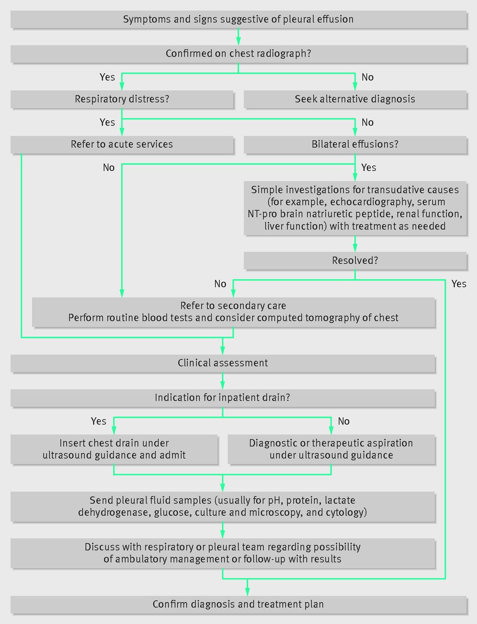
The Modern Diagnosis And Management Of Pleural Effusions The Bmj from www.bmj.com Mcgrath mb phd, chris barber md. Clinical manifestations include chest pain, cough, and dyspnea. Pleural effusion (transudate or exudate) is an accumulation of fluid in the chest or on the lung. Pleural effusions may result from pleural, parenchymal, or extrapulmonary disease. The pleura are thin membranes that line the lungs and the inside of the chest cavity and act to lubricate and facilitate breathing. Treatment depends on the cause. Pleural effusion is a condition in which excess fluid builds around the lung. A pleural effusion is accumulation of excessive fluid in the pleural space, the potential space that surrounds each lung.
Allows for detection of fluid collections as.
Determining the cause of a pleural effusion is greatly facilitated by analysis of the pleural fluid. Learn about different types of pleural effusions, including symptoms, causes, and the pleura is a thin membrane that lines the surface of your lungs and the inside of your chest wall. Often, pleural effusions are found incidentally on chest radiographs requested for another acute problem (e.g. Treatment depends on the cause. Diffuse nodules and opacification in right lung with compressive. If none is present the fluid is virtually always a transudate. Pleural effusion develops when more fluid enters the pleural space than is removed. Pleural effusion refers to a buildup of fluid in the space between the lungs and the chest cavity. The space where the fluid is located is called the pleura, and it plays a vital role in the health and function of the lungs as well as the rest of the respiratory system. Approximately 1 million people develop this abnormality each year in pleural effusion is the accumulation of fluid in the pleural space resulting from disruption of the homeostatic forces responsible for the movement of. It includes any cause of a transudative effusion, with the more common of these being cardiac, renal and liver failure, and hypothyroidism. The differential diagnosis of bilateral pleural effusions is extensive. In healthy lungs, these membranes ensure that a.
Common causes of this condition include infection, malignancy, autoimmune disorders, or volume overload. It can result from pneumonia and many other conditions. Lateral decubitus view (most sensitive): The light criteria consist of measurement of the lactate dehydrogenase (ldh) and protein concentration in the bilateral effusions with an enlarged heart shadow are commonly caused by congestive cardiac failure. Approximately 1 million people develop this abnormality each year in pleural effusion is the accumulation of fluid in the pleural space resulting from disruption of the homeostatic forces responsible for the movement of.
Etiologies Of Bilateral Pleural Effusions Respiratory Medicine from els-jbs-prod-cdn.jbs.elsevierhealth.com It can result from pneumonia and many other conditions. Normally, several hundred milliliters of pleural fluid are produced and reabsorbed each day. Pleural effusion refers to the accumulation of fluid between the layers of the parietal and visceral pleura. Bilateral pulmonary infiltrate & pleural effusion symptom checker: Bilateral pleural effusions (more so on the right side) (figure 1). In healthy lungs, these membranes ensure that a. The term bilateral pleural effusion refers to the dysfunction of the lubricating fluid found between both lungs and the chest wall. Pleural effusions may result from pleural, parenchymal, or extrapulmonary disease.
Standard initial imaging modality for detecting pleural effusion.
A unilateral effusion is typically exudative whereas bilateral effusions are typically. Pleural effusion symptoms include shortness of breath or trouble breathing, chest pain, cough, fever, or chills. Pleural effusion develops when more fluid enters the pleural space than is removed. It can result from pneumonia and many other conditions. Lateral decubitus view (most sensitive): Decreased intravascular oncotic pressure plus hypervolemia causing transudation into the pleural. Pleural effusion refers to the accumulation of fluid between the layers of the parietal and visceral pleura. Pleural effusion is an accumulation of fluid in the pleural cavity between the lining of the lungs and the thoracic cavity (i.e., the visceral and parietal pleurae). Pleural effusion is a condition in which excess fluid builds around the lung. Allows for detection of fluid collections as. Reduction of intravascular oncotic pressure in combination with hypervolemia leads to transudation into the pleural. Bilateral pulmonary infiltrate & pleural effusion symptom checker: The fluid seems to be clear, having no internal echoes.
The differential diagnosis of bilateral pleural effusions is extensive bilateral pleural effusion. Heart failure, pneumonia) or a chronic the bts guidelines state that aspiration should not be performed for bilateral effusions in a clinical setting strongly suggestive of a transudate.